I know that a brain MRI image to most people, might most possibility look like this
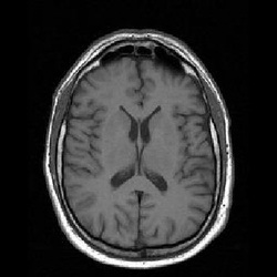
This type of image contrast is often referred to as T1-weighted image (bright white matter, grey cortex, and dark cerebrospinal fluid or CSF). Often, in the science fiction movies, you will see with the main character being bounded on the table (i.e. if u still remember Source Code), an MRI of the character is being traversed up and down, along with rotating motions. The typical plot will be the scientists try to input and/or extract information from our heroes... unfortunately, with the image look like above...rarely much can be done, and in the boring reality, most of the valuable information is not obtained from such image contrast.
There are various mechanism to achieve different contrasts, or in another word, to alter the way different brain tissues look like in an MRI image. You might have the option to highlight structures, such as the tumor or blood vessel; you can also have the liberty to absolutely erase the tissues from an image (if you are interested, check out inversion recovery images). All these contrasts are achieved through manipulating the flip-angle (α), echo time (TE), and repetition time (TR) for determining the turn on and off of the magnetic fields (of course, there can also be the inversion time for the inversion recovery images, and maybe even some other parameters).
To make a simple demonstration of how it works, I decided to use a relatively simple, but widely used MRI method, called the gradient-echo images. This is also used in the "mind-reading MRI". You can predict the image contrast based on the signal equation below:.
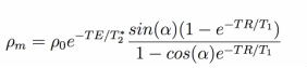
where T1, T2*, and ρ0 are the intrinsic properties of different tissues; TE, TR, and α are the parameters we want to manipulate.
First, let's take a look at the case when TR and TE are fixed, what will happen if we alter the flip-angle.
First, let's take a look at the case when TR and TE are fixed, what will happen if we alter the flip-angle.
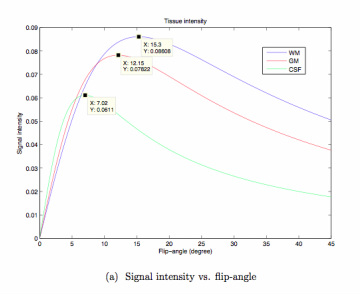
Notice that the signal strength of different tissues attend their own peaks at different flip-angle? At certain flip-angle, the signal strength of two tissues might even overlap. This creates a base for some interesting applications, such as for tissue classification or derivation of the intrinsic properties of bodily tissues.
Now to make the picture prettier, let's throw in another parameter, the echo time (TE) as an additional variable, and still keep the TR unchanged (for our demonstration, TE<TR).
Now to make the picture prettier, let's throw in another parameter, the echo time (TE) as an additional variable, and still keep the TR unchanged (for our demonstration, TE<TR).
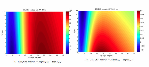
The image above shows the color-coded contrast between white matter and grey matter, grey matter and CSF. The red color means high magnitude and the blue color means low magnitude. If you are bewildered by this demonstration, think about an iso-contour map that marks the lines with the same latitude of a mountain region. With this map, you can easily locate the appropriate TE and flip-angle for the contrast you desire, and you probably notice now that in an MRI it is possible that white matter can indeed look darker than or the same as the grey matter.
One last note, the only thing we didn't alter is TR, which is often related to how long the MRI time could be... As a rule of thumb, if you want to keep a relatively constant image contrast (mostly for the case of T1 images), your flip-angle should increase/decrease with your TR, and this scaling factor is proportional to the square root of (TR-new/TR-old).
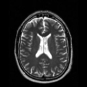
The image below shows a T2-weighted MRI that provides probably more information for diagnostic purposes.
One last note, the only thing we didn't alter is TR, which is often related to how long the MRI time could be... As a rule of thumb, if you want to keep a relatively constant image contrast (mostly for the case of T1 images), your flip-angle should increase/decrease with your TR, and this scaling factor is proportional to the square root of (TR-new/TR-old).
The image below shows a T2-weighted MRI that provides probably more information for diagnostic purposes.


 RSS Feed
