For the longest time, our ancestors all over the world have always believed that the central control unit of our bodies is the beating heart. I suppose whenever there is crucial emotional roller-coaster, heart rate is often the first thing we notice despite the poor brain is working seriously hard to regulate the entire body to adjust to sudden changes. Well..the traditional definition of death using pulse and heart beat may have also played a major part in the old belief.
When we started to realize that the lump of meat sitting in our skull is the real control centre for all our daily activities, many attempts have been made in the past to provide a comprehensive map for navigating this vital structure of ours. One of the earliest, and personally, the most intriguing one, is from the "science" of Phrenology developed by the German physician Franz Joseph Gall in 1796. It is a method of measuring the skulls in relation to the brain regions underneath the skull territory that are responsible for specific functions or characters of the human being. Although today, we might more or less put the sculpture of Phrenology head (yes, I have one in my living room as well) in the oddity section together perhaps with vodoo dolls and electrical therapy chairs, it did provide some preliminary directions on the separation of brain regions and functions. In the modern world, the general divisions of the brain lobes are also named after the skull bones they are under. However, deep under the cortex, there is a whole lot of world going on carrying relay signals between different sub-regions, which will require much more in-depth investigation.
When we started to realize that the lump of meat sitting in our skull is the real control centre for all our daily activities, many attempts have been made in the past to provide a comprehensive map for navigating this vital structure of ours. One of the earliest, and personally, the most intriguing one, is from the "science" of Phrenology developed by the German physician Franz Joseph Gall in 1796. It is a method of measuring the skulls in relation to the brain regions underneath the skull territory that are responsible for specific functions or characters of the human being. Although today, we might more or less put the sculpture of Phrenology head (yes, I have one in my living room as well) in the oddity section together perhaps with vodoo dolls and electrical therapy chairs, it did provide some preliminary directions on the separation of brain regions and functions. In the modern world, the general divisions of the brain lobes are also named after the skull bones they are under. However, deep under the cortex, there is a whole lot of world going on carrying relay signals between different sub-regions, which will require much more in-depth investigation.
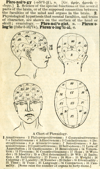
A definition of phrenology with chart from Webster's Academic Dictionary, circa 1895 (image source: wikipedia)
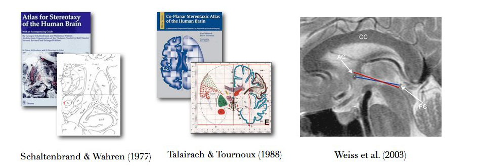
For most people who have studied the human brain in greater details besides the general brain structure divisions, the more elaborate and detailed brain maps, such as the Talairach atlas and/or Schaltenbrand atlas would probably ring a bell. These brain maps are based on the more advanced tissue staining technology and arduous sketches of the European physicians. The general idea here is that the brain is cut into thin slices, and each slice is chemically stained, and outlined for each shown structure. Later on, a few variants of these two atlases were published as well, but the general names are still kept the same. For both of them, the establishment of the coordinate system for the brain are based on two important brain structures, the anterior (AC) and posterior commissures (PC), which, during the time before the invention of MRI, were important landmarks in CT images for the early generation of neuroscientists and surgeons to navigate the brain. For the historical reasons, the habit of using these two structures to define brain map coordinate remains until today even more advanced imaging and computer tools have become increasingly available. With the two landmarks and according to the rule of symmetry, the brain is then investigated under a grid system, or often referred to as "stereotactic space". For many deep brain structures, the idea (and the wishful thinking) is that for all human brains, without slicing open the living brain, the location of the structure is a fixed or scale-up distance in 3D walking from the landmarks. This will help tremendously in the operations as we know more and more about the "functional surgeries". Despite the similar attempts to define the coordinate system in both atlases, one should always be warned that the central lines connecting the two commissure points, which define the anterior-posterior direction in the "stereotactic space", are not exactly the same. For the Talairach atlas, the line connects the superior tip of the anterior commissure and the inferior tip of the posterior commissure, while for the Schaltendbran atlas, the line connects the centroids of these two structures. Although the differences can be rather minute, they change quite much how the brain is oriented in the drawings, and thus the way you navigate yourself in the brain when using these brain maps. I have seen quite a few not-so-careful physicians mixing them up. Also, the brain donor for Talairach atlas is an old French lady. Since one brain cannot be sliced in different directions twice, some sketches were estimated from the slicing directions. However, for Schaltenbrand atlas, there are 3 different brain donors for each direction of slicing. As a result, the measurement may not be very consistent across different slicing directions.
Unfortunately, as you may have guessed, everyone's brain is different, and the generalization that everyone's brain is highly similar and can be navigated by the grid system from these two atlases may not be ideal, particularly when different diseases and aging process can impact the shape of the brain structures. And very importantly...it is still just a piece of drawing! How can this adapt to the new 3D MRI?
While you can now buy a 3D digitized ensemble of Talairach and Schaltenbrand atlases (ask your nearby medical school's library) for your professional use or purely out of curiosity, there are many other versions of more modern digitized 3D brain maps. They are often color-coded and stored as formats that can be directly morphed to match the individual brain anatomy. With the better and stronger MRI scanners, these atlases will often accompany their MRI templates. The most famous ones in the brain imaging field are the MNI305 atlas (http://www.bic.mni.mcgill.ca/ServicesAtlases/MNI305) and ICBM152 atlas (http://www.bic.mni.mcgill.ca/ServicesAtlases/ICBM152NLin2009). The early versions of these atlases were manually aligned to fit the grid system of the Talairach atlas with AC-PC line aligned. The later versions applied more sophisticated image processing techniques to make multiple brain MRIs into one that has the averaged shape of a large population. I have also made my humble share of brain maps for the Parkinson's disease population. If you interested, you may check them out here.
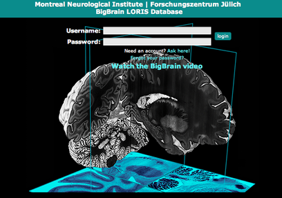
One latest example of the brain map is the project called "Big Brain" developed by the team of Dr. Alan Evans at Montreal Neurological Institute and their collaborators. It was a hyped version of the previous works with more advanced technology. You may zoom-in for incredible details of the brain till the cellular level. The registration is free for use. You can give it a try here. It is mostly for educational purposes so far, application in clinical use is still limited.
From almost superstitious set-up to the cellular view of the human brain map, it was quite a big journey, right? As we add more and more technology and discoveries into the map, the information will only become richer and richer. What would you expect the next map to be like?
While you can now buy a 3D digitized ensemble of Talairach and Schaltenbrand atlases (ask your nearby medical school's library) for your professional use or purely out of curiosity, there are many other versions of more modern digitized 3D brain maps. They are often color-coded and stored as formats that can be directly morphed to match the individual brain anatomy. With the better and stronger MRI scanners, these atlases will often accompany their MRI templates. The most famous ones in the brain imaging field are the MNI305 atlas (http://www.bic.mni.mcgill.ca/ServicesAtlases/MNI305) and ICBM152 atlas (http://www.bic.mni.mcgill.ca/ServicesAtlases/ICBM152NLin2009). The early versions of these atlases were manually aligned to fit the grid system of the Talairach atlas with AC-PC line aligned. The later versions applied more sophisticated image processing techniques to make multiple brain MRIs into one that has the averaged shape of a large population. I have also made my humble share of brain maps for the Parkinson's disease population. If you interested, you may check them out here.
One latest example of the brain map is the project called "Big Brain" developed by the team of Dr. Alan Evans at Montreal Neurological Institute and their collaborators. It was a hyped version of the previous works with more advanced technology. You may zoom-in for incredible details of the brain till the cellular level. The registration is free for use. You can give it a try here. It is mostly for educational purposes so far, application in clinical use is still limited.
From almost superstitious set-up to the cellular view of the human brain map, it was quite a big journey, right? As we add more and more technology and discoveries into the map, the information will only become richer and richer. What would you expect the next map to be like?


 RSS Feed
